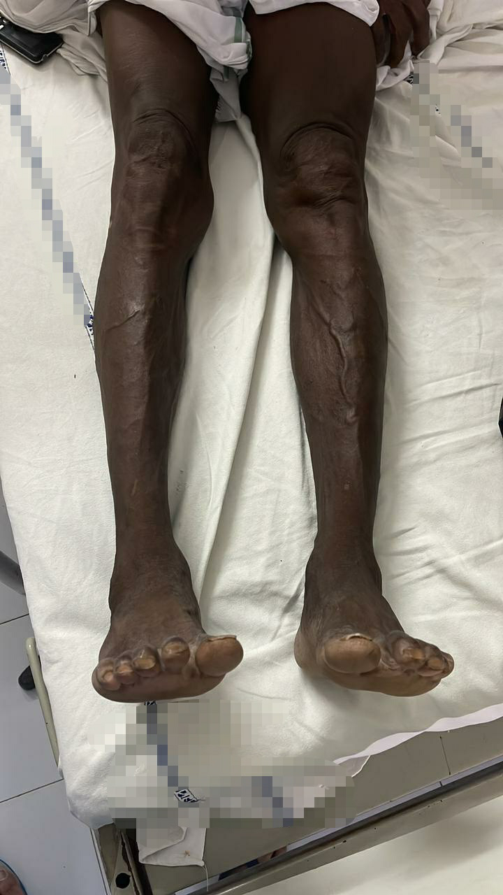
GENERAL MEDICINE CASE 6
"This is an online E log book to discuss our patient's de-identified health data shared after taking his/her/guardian's signed informed consent.
Here we discuss our individual patient's problems through series of inputs from available global online community of experts with an aim to solve those patient's clinical problems with collective current best evidence based inputs.
This E log book also reflects my patient-centered online learning portfolio and your valuable inputs on the comment box is welcome."
Case: A 45 yr old female patient came to opd with b/l lower limb weakness.
I've been given this case to solve in an attempt to understand the topic of "patient clinical data analysis" to develop my competency in reading and comprehending clinical data including history, clinical findings, investigations and come up with a diagnosis and treatment plan.
You can find the entire real patient clinical problem in this link here..
The problems in order of priority I found are
1) B/l lower limb weakness for past 5 months
2) Headache for past 2years
3)multiple body pains,disturbed sleep,
4)Auditory hallucinations ,double vision.
Other complaints
Diagnosed as depression without psychotic features
She was on Bulotin , Esihan's plus for 1 week
Explanation of above complaints in detail.
1-WEAKNESS OF LOWERLIMBS AND SWAYING
Onset:sudden in onset
Duration :since 5 months
Associated conditions :
-NO Tingling and numbness
-history of sudden fall and slurring of speech for few days associated with swaying.
Swaying:
Complaints of swaying to right/left side unable to get up from bed, giddiness on lying down, standing, walking.
mostly associated with cerebellum,may be posterior cerebellar artery ischemia.
Possible diagnosis:
1)A sudden fall may be indicative of vascular cause probably due to stroke or ischemia or infarcts in the brain.
2)Weakness may be caused by any one of the following related to vascular system,muscle or nervous system.
If Artery related
As in case of peripheral artery disease and varicose veins there will be pain,leg skin atrophy,and claudication,telengectasia. As there are no signs of this artery are ruled out.
#if any blood vessel in the brain undergoes infarction then there is a chance of sudden stroke and paralysis of limbs.
If suspecting nervous system related?
May be it is related to nerves because from the history there is history of weakness tingling and numbers in the feet
To understand the nerve related issue A thorough CNS EXamination is done and following are results.
CNS-EXAMINATION
CNS Examination
Higher mental functions-
Consciousness present
Orientation to time ,place and person present
Memory -immediate ,recent and remote present
Speech and emotions -intact
Cranial nerves -
Facial nerve-effected on left side
Motor System-
Tone -hypotonia present in rt upper and lower limbs
Power -
UL . Rt. Lt
-4/5. -4/5
LL
5/5. 5/5
Gait - ataxic gait
Reflexes -
Superficial reflexes - abdominal reflex - absent
Plantar extensor reflex present
Deep reflexes - exaggerated
1.ANATOMICAL LOCATION OF THE PROBLEM
From the above CNS examination findings we can anatomically localise it as UMN LESION because there is hyperreflexia(exaggerated reflexes).
Specific localisation.
Now according to the facial nerve damage the umn lesion is confirmed as above the level of pons.
That is we have to find the exact location of lesion by rule out method.
Possible lesions may be at spinalcord,brainstem,cerebral cortex,thalamus,internal capsule etc.
I think it may be in the cortex region because lower limbs are more affected than upper limbs according to homunculus.
INVESTIGATIONS DONE:
According to colour Doppler test there is soft plaque in the left carotid bifurcation
MRI says that ACA,MCA,PCA are normal so there is no vasculitis cause.
MRI differential diagnosis
1)NEUROMYLITIS OPTICA
2)Multiple sclerosis
3)vasculitis
4)Neurosarcoidosis
5)Neuro-behcets disease
1- MULTIPLE SCLEROSIS (demyelination disorder)
we can rule out this as there is no horse shoe ring enhancement and Dawson fingers and also plaques ,and no triradiate hyperintensity in centre of ponsin MRI .
Plaques can occur anywhere in the central nervous system. They are typically ovoid in shape and perivenular in distribution
2- vasculitis-
we can rule out this also because all the ACA MCA PCA arteries are normal in MRI and also clinically there is no sudden onset of paralysis.
3-neuromylitis optica
Neuromyelitis optica, also called NMO or Devic's disease, is a rare yet severe demyelinating autoimmune inflammatory process affecting the central nervous system. It specifically affects the myelin, which is the insulation around the nerves. NMO mainly affects the spinal cord and the optic nerves -- the nerves that carry signals from the eyes to the brain. As a result, the disease can cause paralysis and blindness. As there are no clinical symptoms above.. we can rule out this.
4-Neurosarcoidosis
The radiographic features of neurosarcoidosis can be thought of as occurring in one or more of five compartments. From superficial to deep they are:
- skull vault involvement
- pachymeningeal involvement
- leptomeningeal involvement (seen in up to 40% of cases 1)
- pituitary and hypothalamic involvement
- cranial nerve involvement
- parenchymal involvement (most common)
- As there is no signs of this we can rule out Neurosarcoidosis.
5-NEURO -BEHCETS DISEASE
Neurological involvement may occur in the course of the disease and have two major forms: vascular and parenchymal . Parenchymal lesions lead to inflammatory lesions in the brain stem, diencephalon, basal ganglia and less frequently, the spinal cord and cerebellum. The cerebral cortices seem to be spared. They usually manifest as bilateral pyramidal signs, unilateral hemiparesis, behavioural changes, sphincter disturbances and headache.
The MRI findings of our case were in keeping with neuro-BD with the classical involvement of brain . These parenchymal lesions are thought to represent vasculitis of small vessels, mainly venular involvement. The differential diagnosis radiologically may include diseases such as neurosarcoidosis, small-vessel vasculitis of brain and viral infections.
Reference link:
Treatment
The treatment of parenchymal neuro-BD consists of glucocorticoids (high-dose pulse intravenous and/or oral) and azathioprine . In refractory cases, anti-TNF alpha therapy with infliximab has shown to be of benefit..!
Present treatment.
1. Ink.Methyprednisolone 1gm in 100 ml NS OD
2. Inj.Zofer 4 mg /i.v /sos
3. Tab.PCM 650mg/PO/sos
4.Tab.Atorvastatin 20 mg/PO to dissolve soft plaques in left carotid bifurcation.
5. Physiotherapy of right upper limb and lower limb
6. BP charting 4 the hrly
THANK YOU.


Comments
My questions on this case:
1)What is the reason for her depression? Is it associated with present condition or she's worried about her inability to continue her routine daily activities or any other social factors?
2) In case if she's not reported to our department and she's from a rural background, where there is no MRI available,is there any other methods to diagnose her based only on her clinical symptoms?
3) How many cases are reported with similar features till now and their survival rate?
4) Without any clinical symptoms like vaginal and oral ulcers how can we confirm it as neuro-behcets disease based only on radiological features?